Family education packRight ankleNot a diagnosisDiscuss with doctors
Mr Sanjeev Chaudhary — Right Ankle Briefing
Calm, precise notes so the family can talk clearly with the treating orthopaedic team at YASHODA Super Speciality Hospitals, Kaushambi.
Age / Sex
42 / Male
Weight
115 kg
Hospital
YASHODA, Kaushambi
Referred by
Dr Akhil Kulshreshtha
X-ray
14 Sep 2026
MRI
15 Sep 2026
Disclaimer: Educational synthesis for family discussion with treating doctors — NOT a diagnosis, NOT a substitute for orthopaedic / infectious-disease judgment. Surgeons often discuss sampling; a needle may be tried first, but tissue is often needed — follow the treating team.
★ 13 Oct 2026 update — new MRI + blood tests
Disclaimer: Educational synthesis for family discussion with treating doctors — NOT a diagnosis, NOT a substitute for orthopaedic / infectious-disease judgment. The two MRIs were done at different centres and read by different radiologists, so differences may reflect interpretation, not real change.
Verdict: broadly STABLE, cannot call it improving
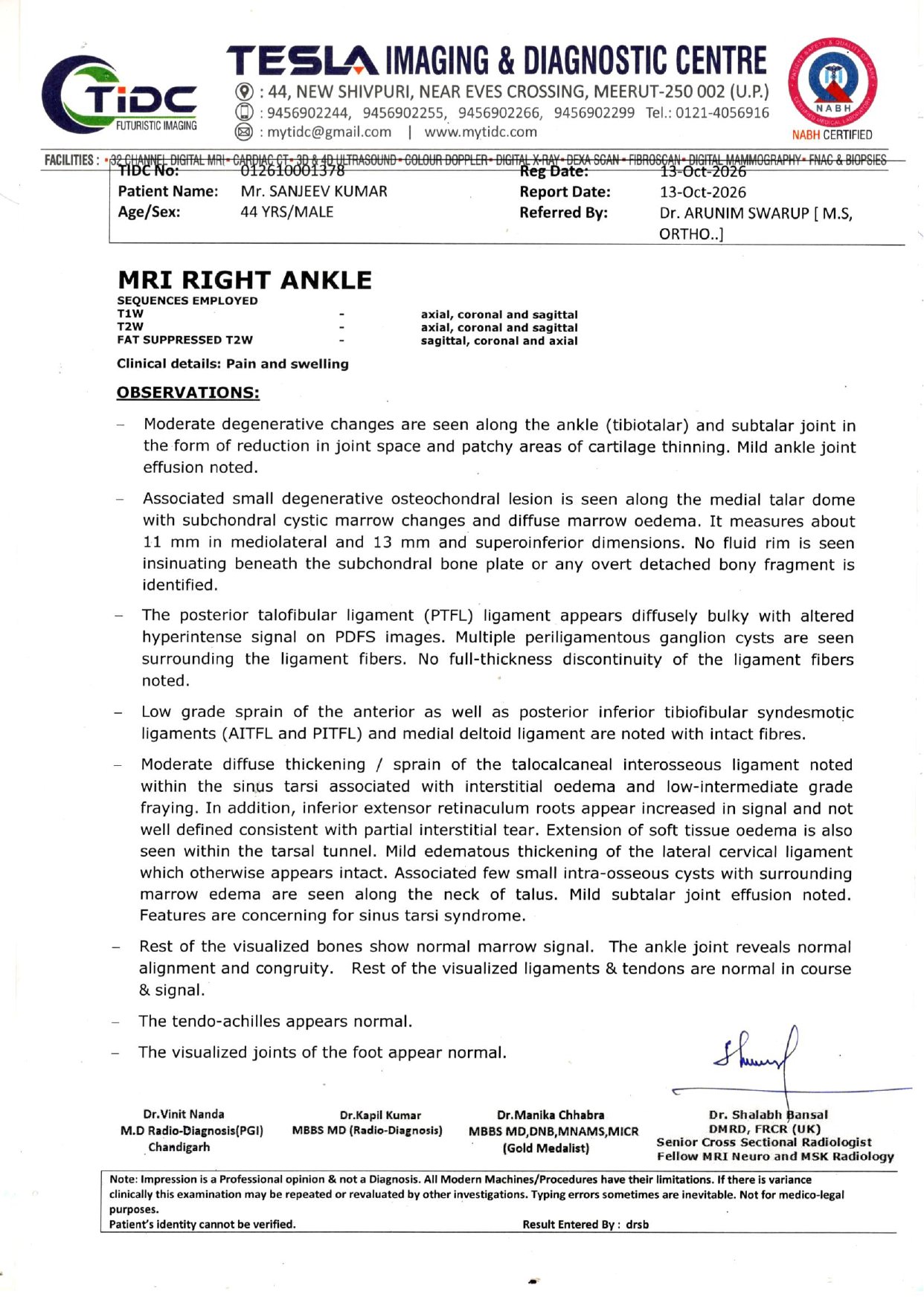
The main talar dome lesion is the same size (13×11 mm in Sep vs 11×13 mm now). Some September "active" features are not described now, but a different reader at a different centre makes that unreliable as proof of healing.
The two MRI reports DISAGREE
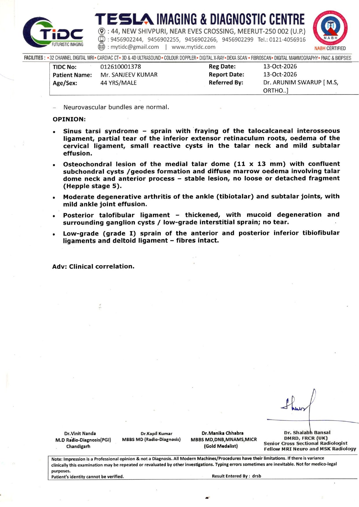
Sep (Yashoda): "infective arthritis – ? tubercular". Oct (Tesla, Meerut): degenerative arthritis + stable osteochondral lesion + sinus tarsi syndrome, with no mention of infection or TB. Doctors must settle this before any biopsy or surgery decision.
Blood: mild inflammation, no TB test done
CRP 11.7 mg/L (normal 0–6) = mild, non-specific inflammation. ESR and TB blood tests (IGRA/Mantoux) were not done. RA factor, uric acid, vitamin D, calcium are not abnormal in a way that explains the ankle.
MRI comparison — 15 Sep vs 13 Oct 2026
Feature
15 Sep 2026 (Yashoda, Dr Mamta Motla)
13 Oct 2026 (Tesla Imaging, Meerut, Dr Shalabh Bansal)
Read-across
Talar dome cyst / lesion
Subchondral cysts, dome of talus 13×11 mm
Osteochondral lesion medial talar dome 11×13 mm, confluent subchondral cysts; "stable, no loose fragment (Hepple stage 5)"
Same size → stable
Second cyst, inferior talus
16×10 mm along inferior articular talus
Not measured; "few small intra-osseous cysts" at talar neck
Can't compare — ask radiologist
Marrow oedema
Talus, extending to anterior calcaneum
Diffuse talar oedema (dome, neck, anterior process); calcaneum not mentioned
The new MRI report names "Mr Sanjeev Kumar", 44 yrs (Sept reports: Sanjeev Chaudhary, 42). Confirm it is the right patient before relying on it.
The two radiology opinions conflict (infection/TB vs degenerative). Ask one musculoskeletal radiologist to read both sets of films side by side.
CRP is still raised; inflammation has not settled.
Urgent care if fever, night sweats, weight loss, rapidly increasing swelling/redness, or inability to bear weight.
New reports (13 Oct 2026)
MRI right ankle — Tesla Imaging & Diagnostic Centre, Meerut, TIDC 012610001378, 13/10/2026, Dr Shalabh Bansal; referred by Dr Arunim Swarup (Ortho). Page 1 (observations).
Tap to open full-size original
Page 2 — opinion. (Two identical scans of this report were received.)
Disclaimer: Educational synthesis for family discussion with treating doctors — NOT a diagnosis, NOT a substitute for orthopaedic / infectious-disease judgment. Sampling approach is the treating team’s call.
MRI of the right ankle shows an infection-pattern arthritis, focused mainly on the subtalar joint (between talus and heel bone), with marrow oedema in the talus, oedema reaching the front of the calcaneum, and two subchondral cysts in the talus (about 13×11 mm and 16×10 mm).
There is loculated / septated fluid and mild synovial thickening at the subtalar joint, plus a milder “reactionary” effusion at the tibiotalar (main ankle) joint, and mild soft-tissue swelling around the foot and distal leg.
The plain X-ray a day earlier looks almost normal — bones and joint spaces look fine on film. That often means bone destruction has not yet advanced far enough to show on X-ray, so disease may still be early-to-moderate on the destruction scale, even though symptoms have lasted years.
The radiologist’s impression is: features suggestive of infective arthritis — ? tubercular, and asks for clinical correlation. In India, osteoarticular TB is an important possibility for a long-standing single joint — but MRI alone does not confirm TB.
At 115 kg, every step loads the ankle heavily; weight is a load factor and rehab partner, not a “cause” or a cure by itself.
Family understanding of the plan: doctors will discuss how to get a sample (needle aspiration may be tried first; keyhole or open biopsy often needed for tissue), then possibly surgery to clear infection and address damaged cartilage — that tissue work is how TB vs pyogenic infection is actually proved. See Needle vs cut.
Often means destruction is not yet advanced on plain film.
TB = suspicion only
“? Tubercular” is a leading guess, not confirmation.
★ Original hospital reports
Disclaimer: Scans of the written reports from YASHODA Super Speciality Hospitals, Kaushambi. Tap any image to open full size. Educational pack — not a new diagnosis.
Keep these with the MRI films when you meet the orthopaedic team.
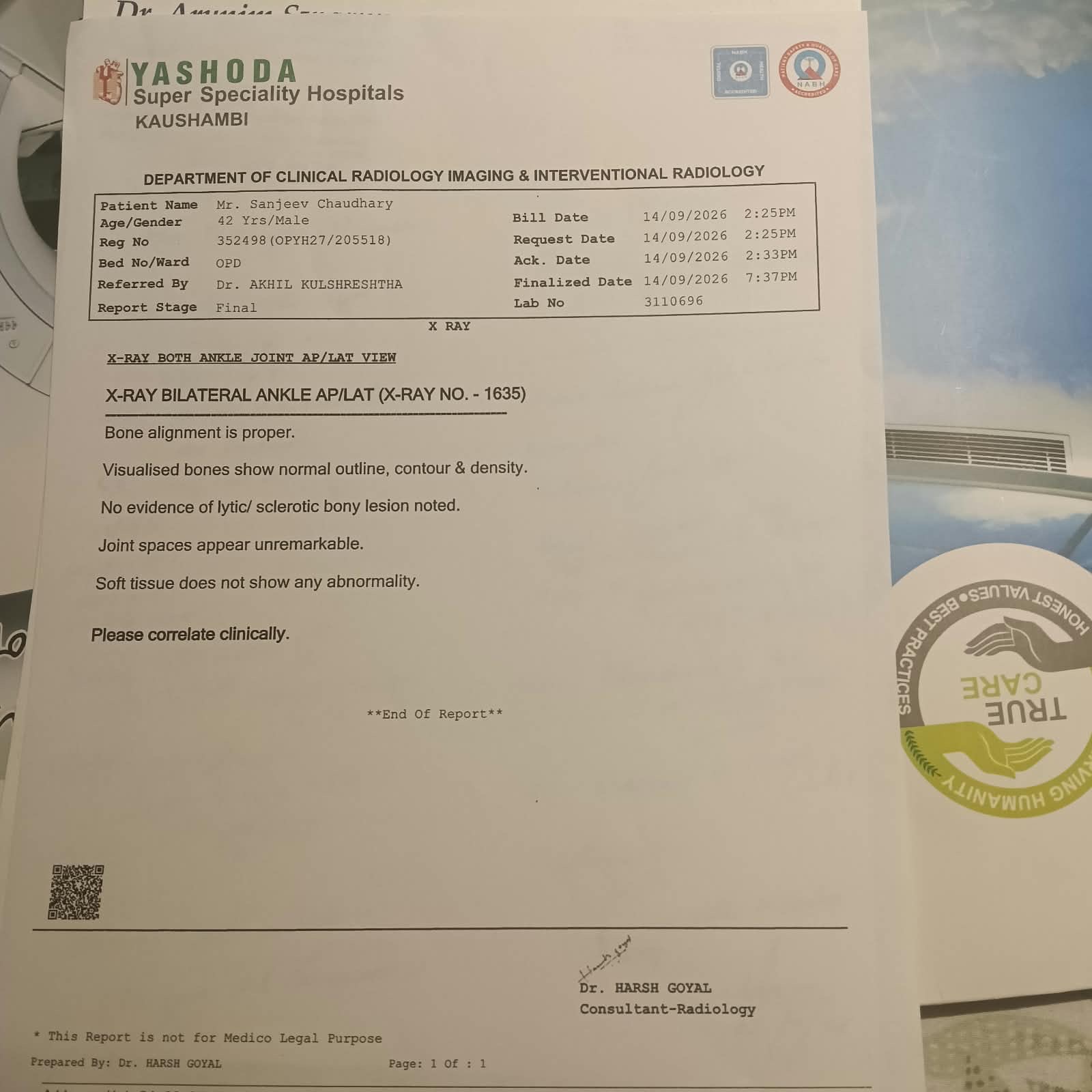
X-ray report — Lab 3110696, 14/09/2026,
Dr Harsh Goyal. Both ankles AP/LAT — essentially normal plain film.
Tap to open full-size original
2 What the MRI found
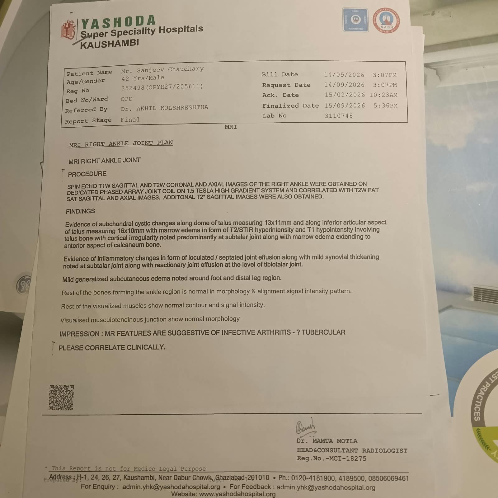
Lab 3110748 · Finalised 15/09/2026 5:36 PM · Dr Mamta Motla (Head & Consultant Radiologist) · Right ankle MRI
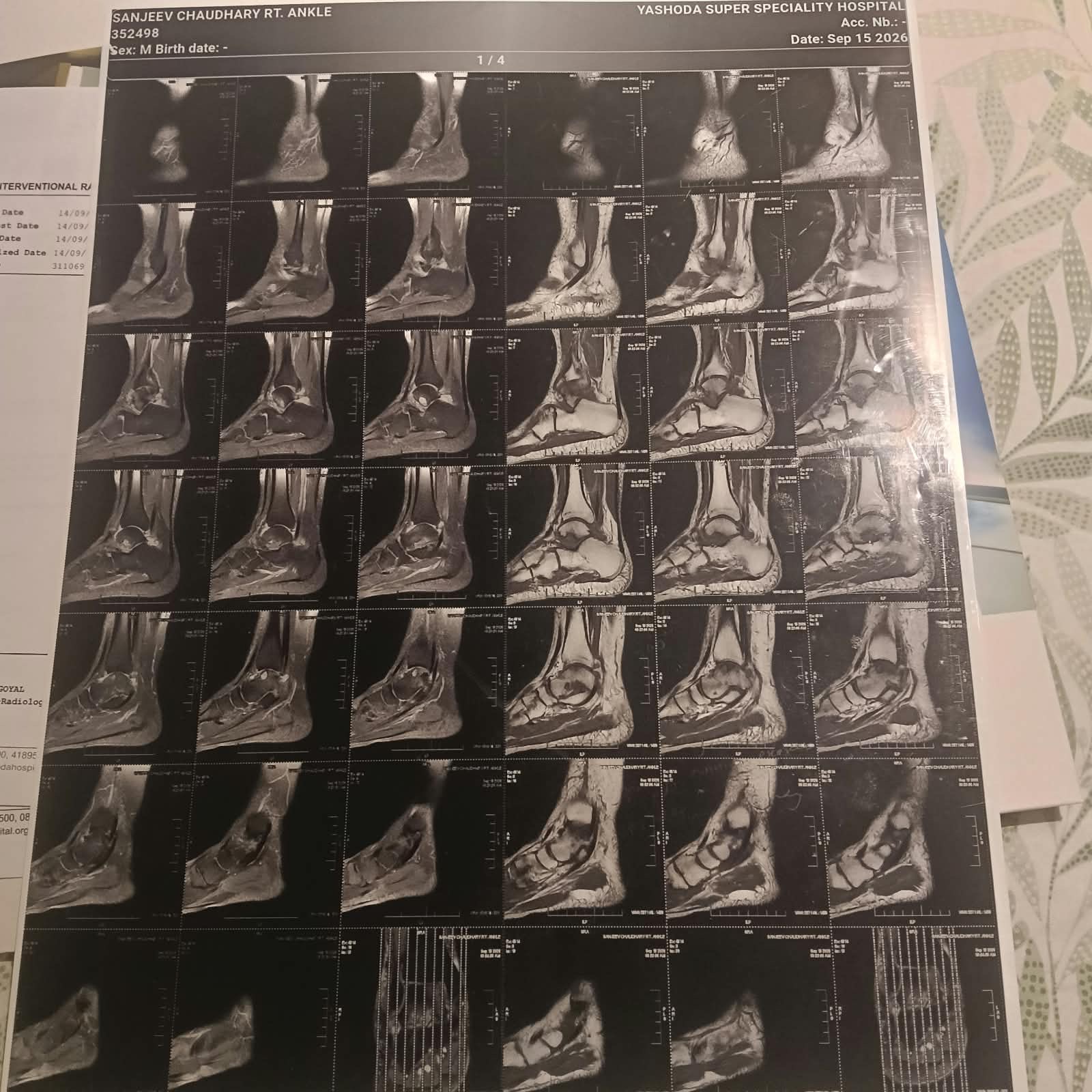
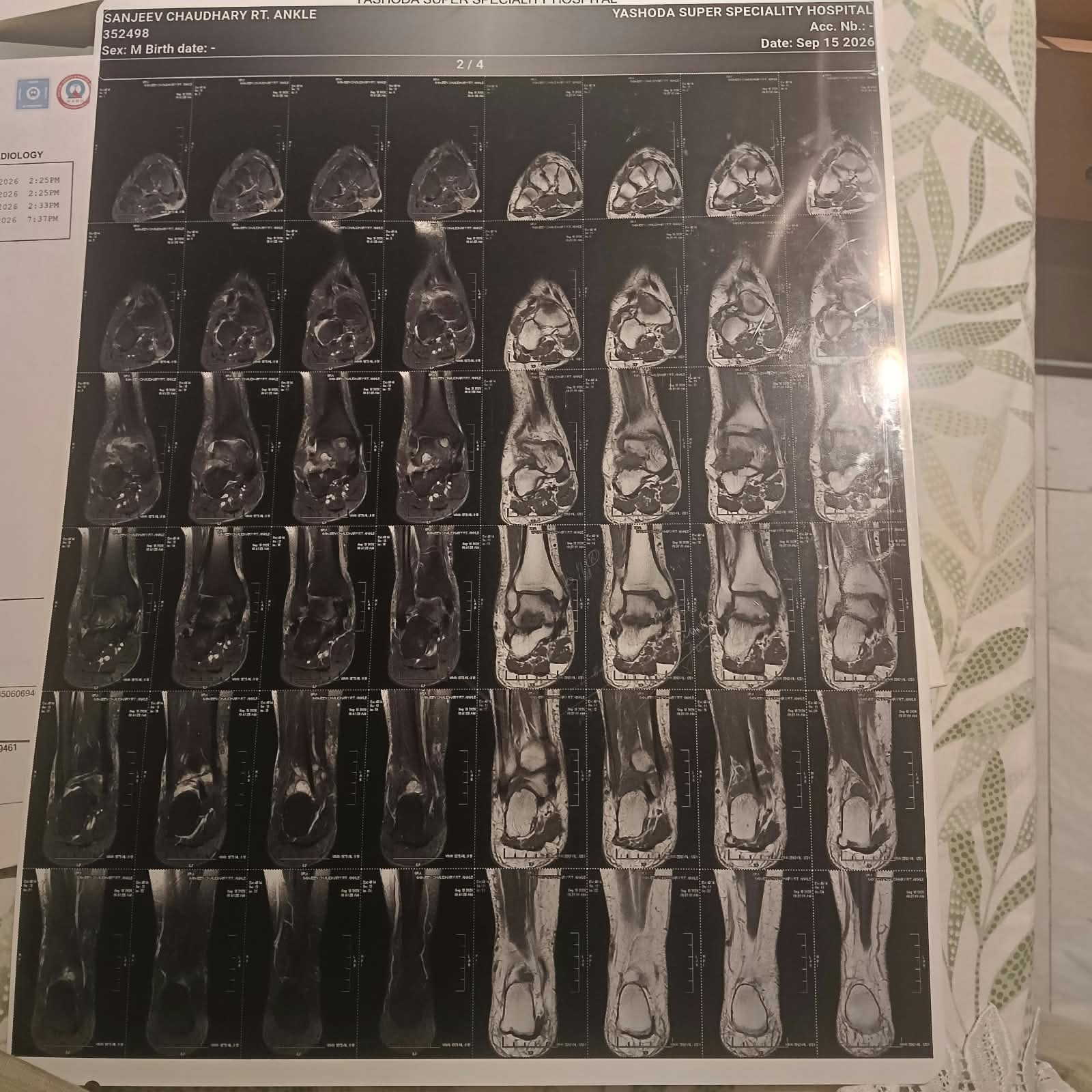
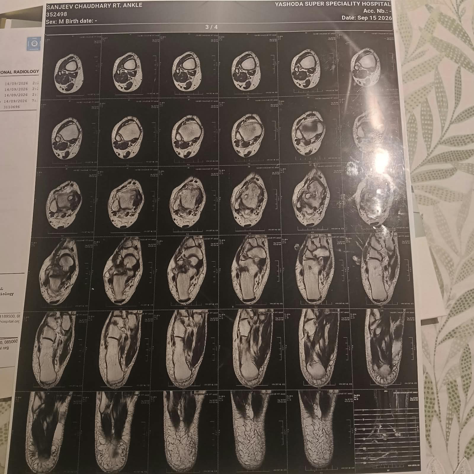
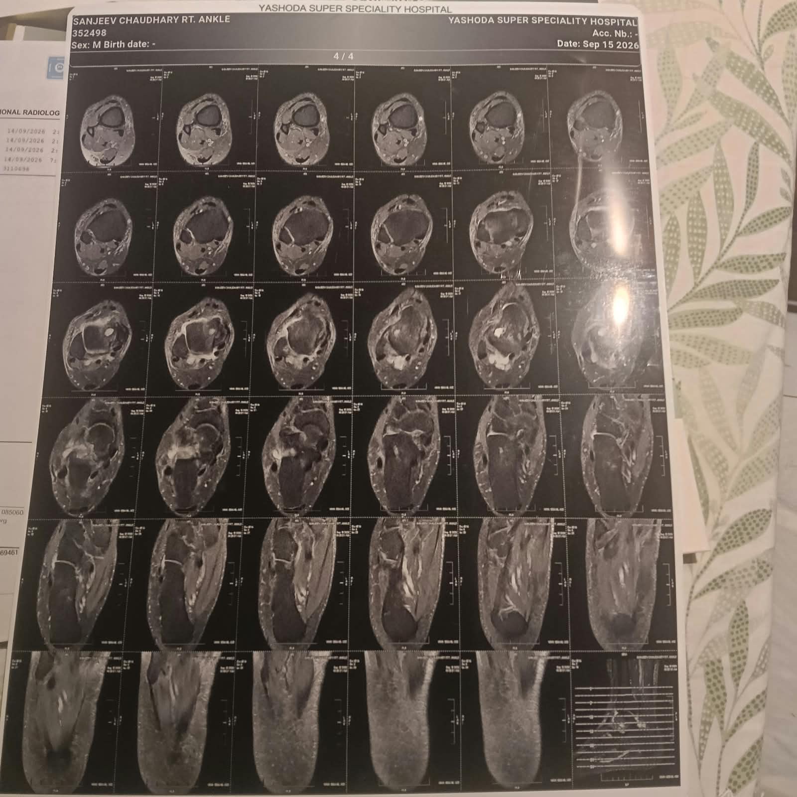
Disclaimer: Educational synthesis for family discussion with treating doctors — NOT a diagnosis. MRI films (4 pages, 15 Sep 2026) are visual context only; do not invent slice-level diagnoses beyond this report.
Official impression (verbatim)MR FEATURES ARE SUGGESTIVE OF INFECTIVE ARTHRITIS - ? TUBERCULAR. PLEASE CORRELATE CLINICALLY.
Key focus
Subtalar joint — oedema + irregularity
Marrow edema (T2/STIR hyper, T1 hypo) involving talus with cortical irregularity predominantly at subtalar joint; oedema extending to anterior calcaneum.
Layman: The “lower” joint under the ankle bone (talus sitting on the heel bone) looks inflamed inside the bone marrow — classic place infection can sit quietly for a long time.
Fluid + lining
Septated effusion + synovial thickening
Loculated/septated joint effusion + mild synovial thickening at subtalar joint.
Layman: Sticky, compartmented fluid and a slightly thickened joint lining — more like inflamed / infected synovium than simple clear fluid from a sprain.
Cysts in talus
Two subchondral cystic changes
Along dome of talus 13×11 mm AND along inferior articular aspect of talus 16×10 mm.
Layman: Small fluid-filled pockets next to the joint surface of the talus — can be seen with infection, arthritis, or cartilage stress; here they sit inside an infection-pattern picture.
Main ankle joint
Tibiotalar reactionary effusion
Reactionary joint effusion at tibiotalar joint.
Layman: Extra fluid in the “upper” ankle (shin bone–talus), described as reactive — often secondary to the hotter process below at the subtalar joint.
Soft tissue
Mild subcutaneous oedema
Mild generalized subcutaneous edema around foot and distal leg.
Layman: Mild waterlogging of soft tissues around the foot and lower leg — matches swelling you can see/feel; not the main driver of the diagnosis.
Reassuring
Other bones & tendons OK
Rest bones/muscles/musculotendinous junctions normal morphology.
Layman: No report of widespread bone collapse or major tendon tears — problem is localised to the described joint/marrow pattern.
Simple teaching diagram — not a scale drawing of the MRI slices. Star = where the report concentrates: subtalar infection-pattern change.
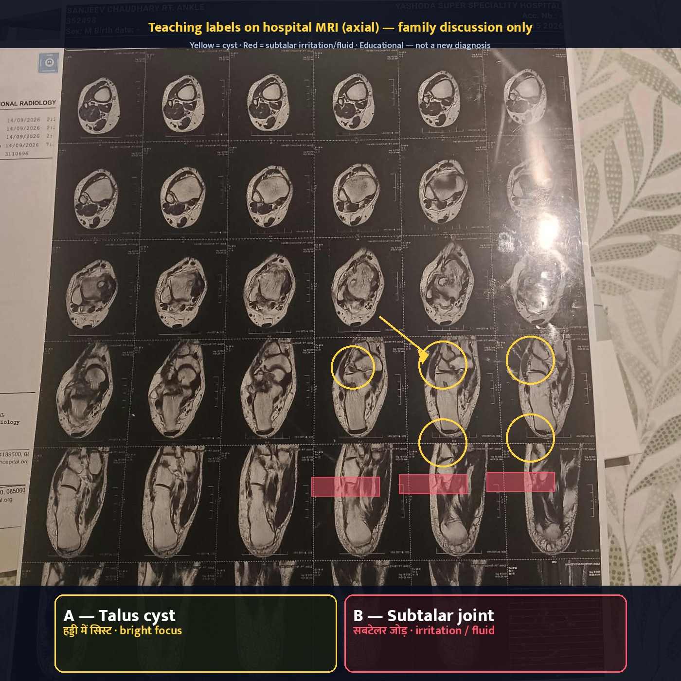
★ MRI films with teaching labels
Disclaimer: Coloured overlays are educational only for family understanding. They are not a new radiology report. Treating doctors decide meaning. MRI impression remains: infective arthritis — ? tubercular (suspicion only).
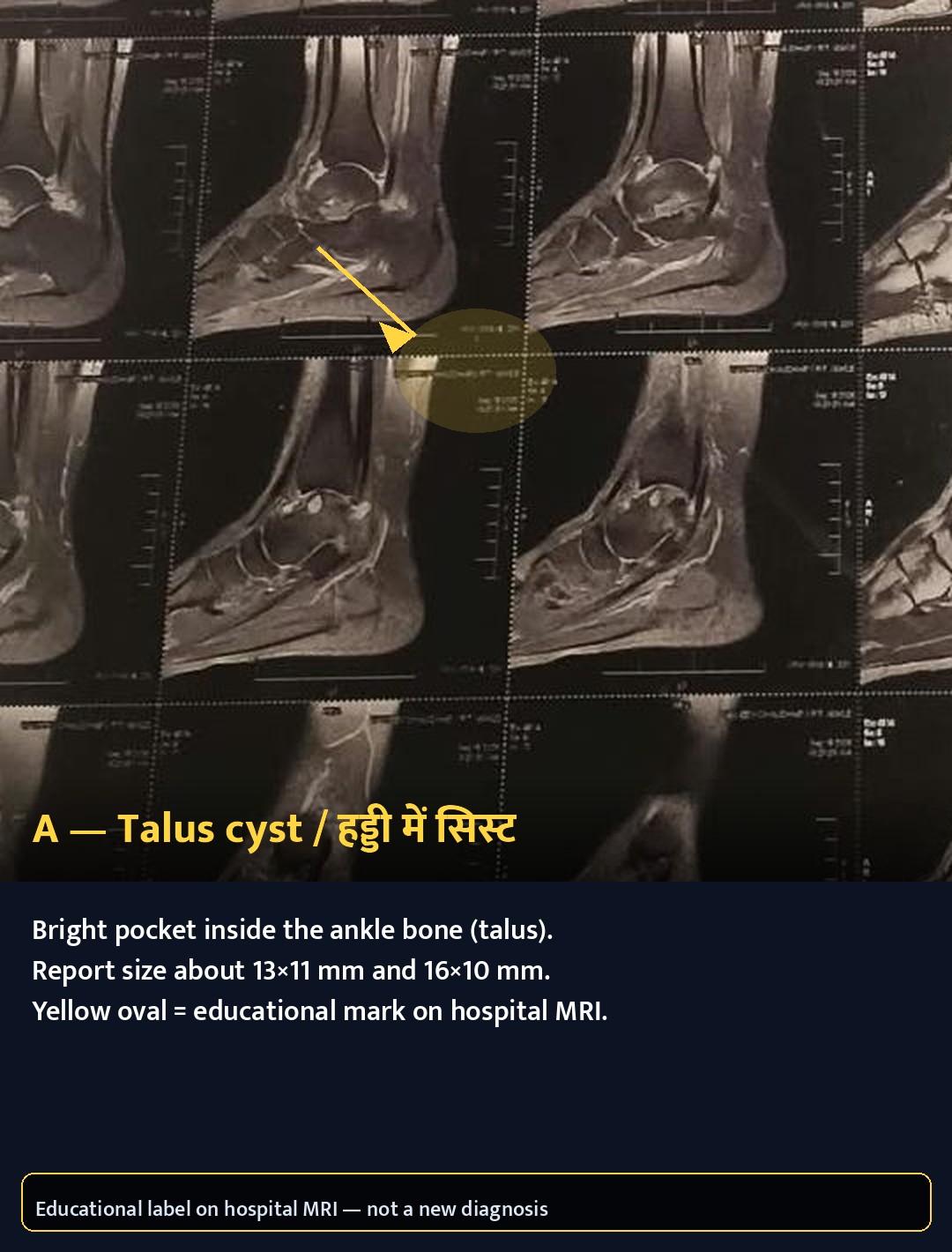
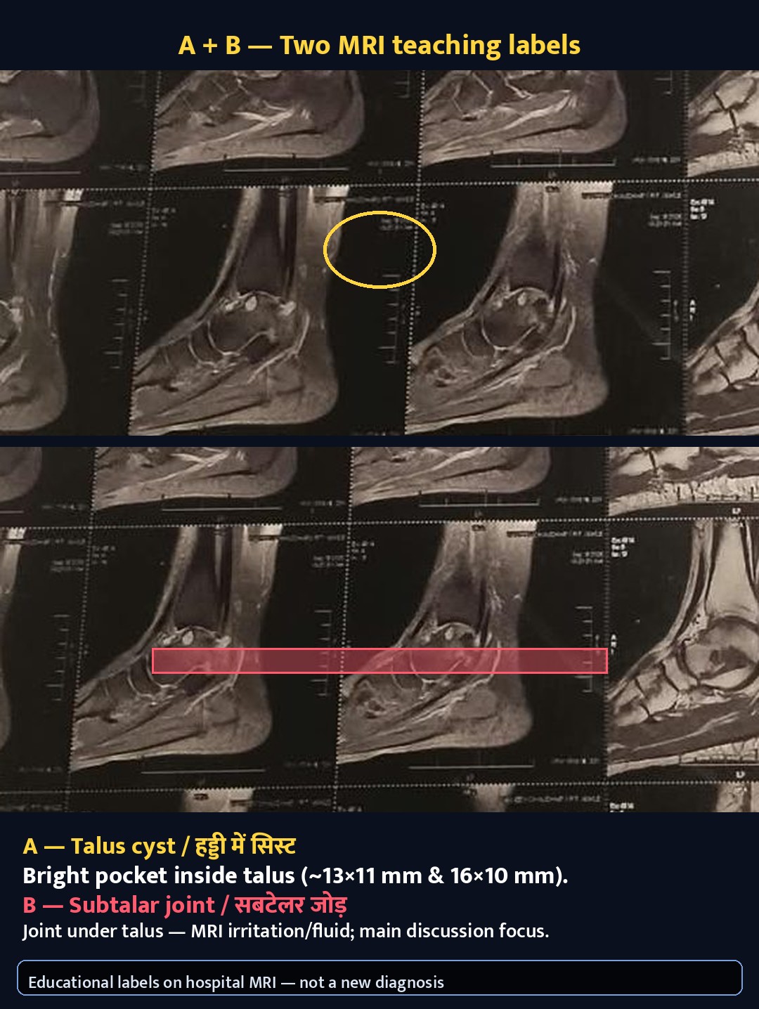
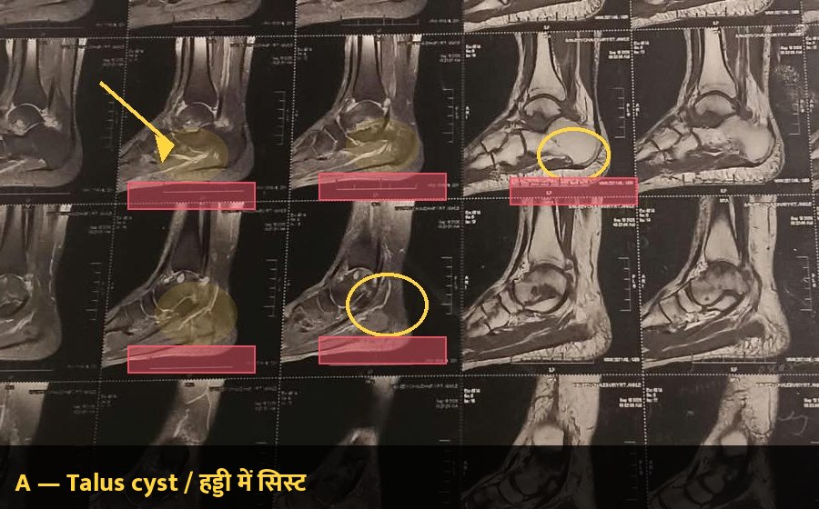
A = Cyst — fluid-filled pocket inside the talus bone (report sizes about 13×11 mm and 16×10 mm).
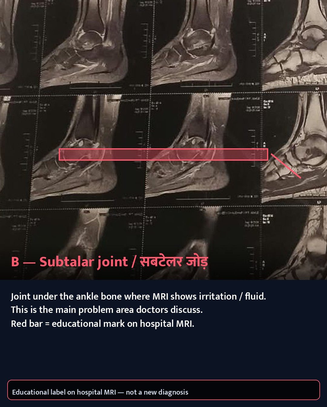
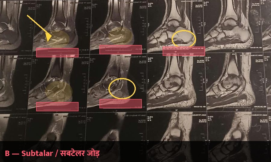
B = Subtalar — the joint under the ankle bone (talus on calcaneus) where MRI shows inflammation/fluid — the main focus.
Phone cards first — large in-frame bilingual labels so you can read without zooming; full sheets are secondary.
Phone teaching cards (start here)
A — Talus cyst / हड्डी में सिस्ट
Bright pocket inside the talus. Report size about 13×11 mm and 16×10 mm.
HTML caption backup: Yellow = cyst. Educational overlay — not a new diagnosis.
Readable on phone without zoom · tap for full size
B — Subtalar joint / सबटेलर जोड़
Joint under the ankle bone — MRI irritation/fluid; main problem area doctors discuss.
HTML caption backup: Red = subtalar. Educational overlay — not a new diagnosis.
Readable on phone without zoom · tap for full size
A + B together — both findings on one phone card (same legend as teach-legend-phone.jpg).
A = talus cyst (yellow) · B = subtalar joint (red).
Full film sheets (secondary — labels in bottom banner)
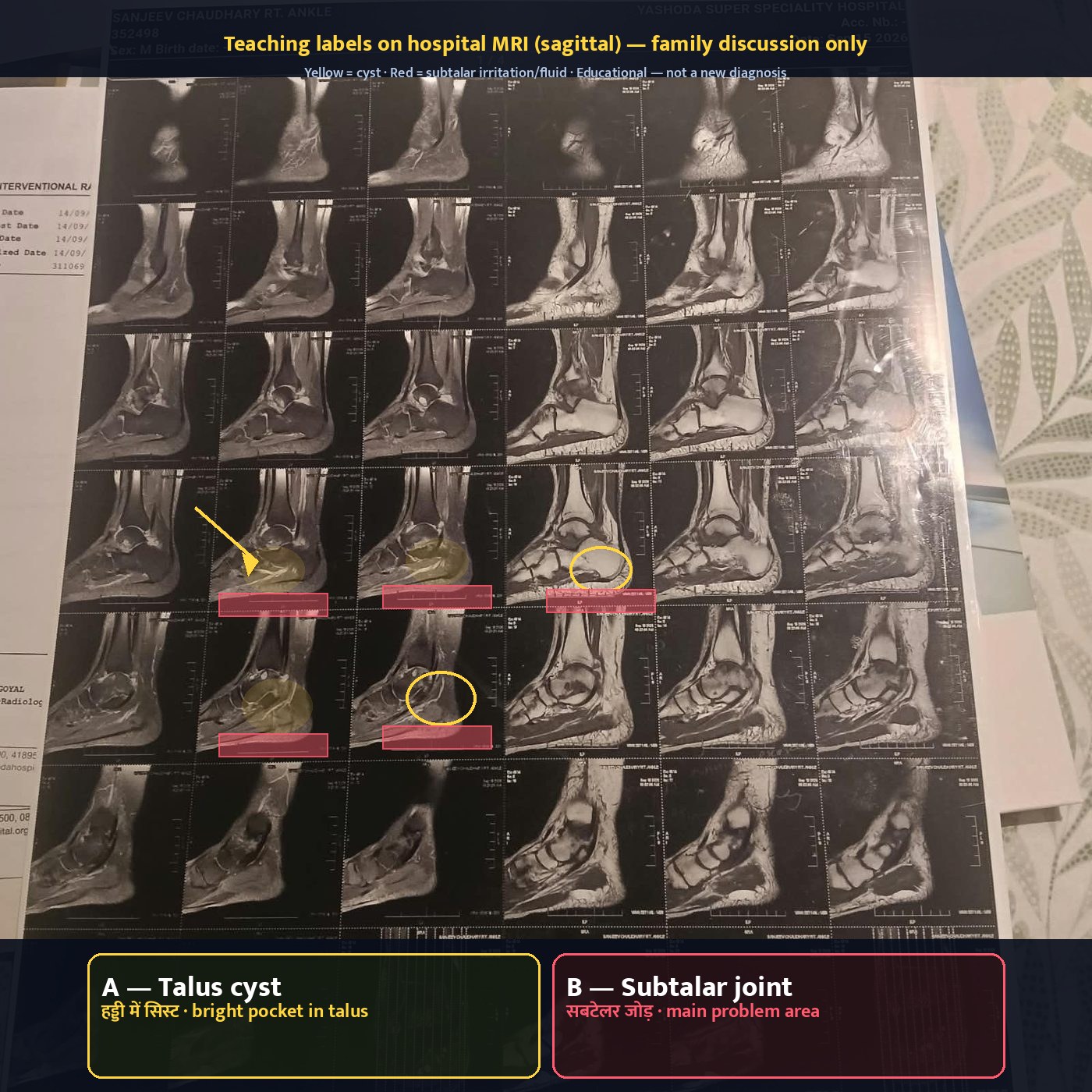
Callout text is in the bottom safe margin (not the right edge), so zooming into slices does not crop A/B explanations.
Film 1/4 (sagittal) — full sheet. Yellow on-slice marks = talus cyst (A); red bars = subtalar (B). Explanations in bottom pills.
MRI impression: infective arthritis — ? tubercular (suspicion only).
Lab 3110696 · Finalised 14/09/2026 7:37 PM · Dr Harsh Goyal · Both ankles AP/LAT
Disclaimer: Educational synthesis for family discussion with treating doctors — NOT a diagnosis, NOT a substitute for orthopaedic / infectious-disease judgment.
What X-ray said
Alignment proper; normal outline/contour/density; no lytic/sclerotic lesion; joint spaces unremarkable; soft tissue no abnormality. Essentially a normal plain film.
Why that still fits
Early osteoarticular TB and early septic arthritis often look quiet on X-ray for weeks–months while MRI already shows marrow oedema, synovitis, and effusion.
Take-home: Normal X-ray does not cancel an abnormal MRI. X-ray is less sensitive for early marrow and soft-tissue infection. MRI is doing the “early warning” job here; tissue sampling does the confirmation job.
X-ray strengths: alignment, big bone loss, obvious fractures.
MRI strengths: marrow oedema, synovitis, loculated fluid, early cartilage/subchondral change.
Classic pattern: chronic monoarticular pain + normal X-ray + “infective” MRI → raise TB/septic work-up, don’t stop at “arthritis of age”.
4 Differential diagnosis (India context)
Disclaimer: Educational synthesis — MRI impression is suspicion, not confirmation of TB. Final label needs clinical exam + labs + tissue. Colour badges = teaching likelihood cues only, not probabilities.
Possibility · likelihood
Typical pattern
How it fits Sanjeev’s picture
Typical next step
Relative recovery / rest burden
Osteoarticular TBAMBER · Leading suspicion on MRI
Not confirmed — “?” only
Commoner in India than West; often one joint; chronic months–years; may have cold abscess; cartilage loss often slower than hot pyogenic infection
Radiologist’s leading “?” — years of pain/swelling, monoarticular ankle, MRI infective pattern
Tissue/fluid: GeneXpert + histo + AFB culture + routine culture; ortho follow-up; ATT only if confirmed/strongly indicated by team
If TB+: often months of ATT + load plan — longest pathway
Pyogenic septic arthritisRED · Must exclude
Usually more acute (days–weeks), fever more common — but chronic smoldering forms exist
Must still be ruled out on the same sample (Gram stain + bacterial culture)
Urgent culture; antibiotics ± drainage per treating team if confirmed
If pyogenic+: antibiotics/drainage — timing set by surgeons/ID
Inflammatory arthritis (RA / seronegative)BLUE · Less typical
Monoarticular years = lower fit
Often multiple joints, morning stiffness, systemic clues
Less typical as only one ankle for years — still discuss blood markers with doctors
Rheumatology path if infection work-up negative / markers point inflammatory
Meds + physio path — not the MRI’s leading story
Advanced OA + cystsGREEN · Low likelihood alone
Mechanical wear, osteophytes, joint-space loss on X-ray
X-ray nearly normal; MRI shows septated effusion + synovitis + infective-pattern oedema — not pure mechanical OA
Does not replace infection work-up; reconstructive talk only after infection controlled if needed
X-ray nearly normal argues against pure advanced OA
Emphasise for family: “Suggestive of infective arthritis — ? tubercular” means this pattern looks like infection, and TB is a top Indian differential. It does not mean “he has proven TB ankle” until tissue/fluid tests say so.
Educational downtime ranges (not prescriptions): needle / day-care usually shortest rest → arthroscopy (keyhole) often weeks → open surgery + long ATT if TB can mean months. Treating team sets the real plan.
★ Expert view — working synthesis
GC_Health educational synthesis for family–doctor discussion. This is NOT a prescription, NOT a radiology re-read, and does NOT replace the Yashoda orthopaedic / ID team. MRI films and reports remain the primary hospital record.
Working hypothesis (unproven)
Infective arthritis focused at the subtalar joint, with marrow change and talus subchondral cysts. In India, osteoarticular TB is an important differential on this MRI pattern — but it is unproven until GeneXpert / culture / histopathology (and clinical correlation) say so. Pyogenic infection must be excluded on the same sample.
Decision tree (family checklist)
Confirm sample plan with ortho — prefer adequate tissue if aspiration volume/quality looks inadequate.
Send the full panel: GeneXpert/CBNAAT + histopathology + AFB/mycobacterial culture + routine bacterial culture/Gram stain.
If TB positive / team starts ATT: medical therapy + orthopaedic follow-up; agree weight-bearing / load plan.
If pyogenic positive: antibiotics and drainage strategy per surgical/ID team.
If inflammatory pathway: rheumatology work-up when infection is reasonably excluded.
Cartilage / reconstructive talk only after the infection question is controlled — not before tissue answers.
Possible solution paths · rough rest burden
Educational ranges for family orientation — actual downtime is set by the treating team and the final diagnosis.
Path 1 — Needle / day-care
Image-guided or clinic aspiration / needle biopsy when fluid/tissue can be obtained safely. Useful first step; may be inconclusive.
Usually shortest downtime
Path 2 — Keyhole (arthroscopy)
Better tissue + washout options when needle is inadequate or surgeons need joint visualisation.
Often weeks of relative rest
Path 3 — Open ± long ATT
Open debridement if required; if TB confirmed, anti-tubercular therapy often runs months with ortho follow-up and load planning.
Longest pathway (months if TB)
Remember: Needle < Keyhole weeks < Open + long ATT months — as a teaching sketch only. Yashoda team chooses the real sequence.
5 Why sample / biopsy / surgery is being discussed
Disclaimer: Surgeons often discuss tissue sampling. A needle may be tried first, but tissue is often needed for osteoarticular TB work-up. This section explains why — it is not a consent form; the treating team decides.
For suspected osteoarticular TB, the gold standard is tissue and/or fluid sent for a full panel — not MRI alone:
GeneXpert / CBNAAT on tissue or pus (rapid TB DNA + rifampicin resistance clue)
AFB smear and mycobacterial culture (culture takes time but remains important)
PLUS routine bacterial culture + Gram stain — so pyogenic septic arthritis is not missed
Aspiration alone
Needle fluid helps, but sometimes volume is low or organisms are scarce — result can be inconclusive.
Arthroscopic / open biopsy
Often needed when fluid alone is not enough. Small portals or an incision let the surgeon see, sample tissue, and often debride infected synovium or pus — diagnostic and sometimes therapeutic. See Needle vs cut.
Why this matters
Wrong empiric treatment delays healing. Right sample guides ATT vs antibiotics vs both, and whether more cartilage work is needed.
Family-reported plan: get a proper sample (needle first if safe, then keyhole/open if tissue needed) → then possibly further surgery for septic arthritis / infection / damaged cartilage. Confirm exact steps, risks, and consent wording with the operating surgeon.
5b Can TB sample be taken with a needle (no big cuts)?
Disclaimer: Family education only. The treating orthopaedic / TB team decides the safest sampling route. This is not a consent form or a prescription.
Short answer: a needle(सुई / aspiration) can sometimes get fluid first — but for ankle/foot TB suspicion, doctors often still need tissue(ऊतक) from a small keyhole or open biopsy.
What the sample is for
MRI saying “? tubercular” is suspicion only. Lab work on fluid/tissue aims for:
Histopathology — granuloma / caseation pattern under the microscope
Mycobacterial culture — slower, still important
AND routine bacterial culture + Gram stain — so pyogenic (ordinary) infection is not missed
Four approaches (least → more invasive)
A. Needle aspiration (arthrocentesis) — usually no stitches; needs enough fluid. Often not enough alone for ankle/foot TB. Negative fluid ≠ no TB.
B. Image-guided needle / core biopsy — still a needle; may sample synovium or bone near the MRI focus.
C. Arthroscopic (keyhole) biopsy(कीहोल) — small portals ~0.5–1 cm; better tissue; sometimes debridement; often 1–2 fine stitches or strips per portal.
D. Open biopsy / debridement — proper incision + stitches when the focus is hard to reach or a bigger clean-out is planned.
Needle
Stitches usually none
Keyhole (arthroscopy)
Stitches usually few small
Open biopsy
Stitches usually more
Caption: stitches — usually none / few small / more, depending on approach. Dark-theme teaching sketch only — not an operative plan.
About the “injection only” hope: It is reasonable to ask whether a safe fluid pocket allows image-guided aspiration first. But osteoarticular TB work-ups often need tissue. Skipping a needed biopsy just to avoid a small cut can delay the correct medicine. Do not start random antibiotics or ATT before the planned sample unless the doctors order it.
Questions for the surgeon (checklist)
Can we try image-guided aspiration first if a safe fluid pocket exists?Least invasive start — only if the team agrees it is useful.
If fluid is taken: send for GeneXpert + mycobacterial culture + routine bacterial culture/Gram?Write all three on the request form.
If dry tap or negative fluid — what is Plan B: arthroscopic tissue vs open?Know the next step before leaving clinic.
Will histopathology be done in the same sitting if keyhole/open?Histo + micro together are stronger.
Will sampling target the subtalar focus (MRI focus), not only the easier tibiotalar joint?Sample the disease — not only the easy joint.
Rest / healing — typical discussion ranges
Note: Typical ranges for family discussion only — NOT a prescription. Your surgeon’s plan wins. At 115 kg, walking limits are often stricter.
Approach
Stay
Wound
Walking (variable; stricter at 115 kg)
Aspiration
Day-care
~1–3 days
Often short relative rest
Image-guided needle
Day-care / overnight
Small wound
Few days limited load if bone sampled
Arthroscopy
Day-care to 1–2 nights
Portals ~1–2 weeks; stitches ~10–14 d if used
Partial / non-weight often weeks
Open
Longer possible
Incision ~10–14+ days
Weeks non-weight common
If TB is confirmed, ATT(anti-TB medicines) often runs many months (osteoarticular typically 9–12 months in Indian practice — team’s call). Wound healing ≠ ATT clock — stitches come out long before medicines finish.
Evidence / sources note (short, honest)
Published orthopaedic / osteoarticular-TB literature and Indian practice summaries prefer tissue over swab/sinus material for OA-TB diagnosis (WAIOT-style guidance and OA-TB reviews).
Ankle arthroscopy series report higher confirmation rates when aspiration alone is inconclusive — keyhole sampling can help after a dry or negative tap.
GeneXpert on tissue speeds TB detection vs culture alone; culture is still needed.
Optional reading pointers (not Sanjeev’s hospital papers): Journal of Orthopaedic Surgery and Research arthroscopic ankle TB series; PMC reviews on the role of surgery in osteoarticular foot/ankle TB. Ask the treating team which local protocol they follow.
6 What to ask the surgeon before consent
Disclaimer: Checklist for empowered discussion — not medical orders. Tick with the treating team.
Will tissue go for GeneXpert / CBNAAT (not only culture)?Ask them to write it on the request form.
Will histopathology (granuloma / caseation) be done on the same sitting?Histo + micro together are stronger than either alone.
Bacterial culture + Gram stain also ordered?Covers pyogenic septic arthritis.
How many portals / incisions? Open vs arthroscopic?Know the scar and approach.
Weight-bearing plan after surgery?Non-weight / partial / walker timeline — critical at 115 kg.
If TB+: ATT duration? (osteoarticular often 9–12 months typical)Confirm with ID / orthopaedic TB protocol they follow.
Chest X-ray / sputum for pulmonary TB screen?Joint TB can travel with or without active lung disease.
Baseline CRP, ESR, CBC, HIV (and other labs they recommend)?Track inflammation and rule out important co-factors.
Is a second opinion from an orthopaedic infection specialist useful before major cartilage work?Reasonable when infection vs inflammatory remains open.
7 Weight 115 kg — load, not blame
Disclaimer: Weight loss is an adjunct, not a cure for suspected infective arthritis. Do not delay biopsy/surgery for a diet plan.
Every step multiplies force through the ankle. At 115 kg (obesity), that load makes pain feel worse, swelling linger, and post-op rehab harder — especially if weight-bearing is restricted.
Before / around surgery
Discuss walker/crutches fit, bed-to-chair transfers, and home support. High load + non-weight-bearing needs a real plan.
Adjunct goals
Steady, doctor-approved weight reduction helps long-term joint comfort and heart/metabolic health — parallel track, not a substitute for infection work-up.
8 Timeline & red flags → urgent care
Disclaimer: Educational only. When in doubt, go to emergency / call the treating doctor.
Seek urgent care if: fever with chills, night sweats that are new/worsening, discharging sinus near the ankle, sudden inability to put foot down / walk, new numbness or foot-drop feeling, rapidly spreading redness, or severe uncontrolled pain.
Chronic background: years of difficulty walking, continuous pain, swelling — matches a smoldering process; still needs planned (not panicked) work-up.
After biopsy/surgery: watch wound discharge, fever, calf swelling (clot risk discussion with surgeons), and medication side-effects if ATT starts.
9 Action pack for family — this week
Disclaimer: Practical family steps — final medical decisions rest with Yashoda treating team.
13 Oct update: Show both MRIs (Sep Yashoda + Oct Tesla) to the treating orthopaedic surgeon and ask one MSK radiologist to compare the films directly: is this infection/TB or degenerative + osteochondral lesion?
Confirm the name on the Oct MRI ("Sanjeev Kumar", 44) is a clerical error for the same patient.
Still-missing tests to ask about: ESR, IGRA (TB blood test) or Mantoux, chest X-ray, HIV, ferritin. Repeat CRP in 4–6 weeks for a trend.
Raised ALT (56.6) and HbA1c 6.3: tell the doctor; these matter if anti-TB drugs or surgery are planned.
Sampling/biopsy decision now depends on resolving the conflicting MRI readings — do not skip it if the team still advises it.
Keep both reports (X-ray 14 Sep + MRI 15 Sep) and the 4-page MRI films together in one folder / phone album.
Book / attend the orthopaedic review with the checklist in section 6 printed or on phone.
Ask explicitly that biopsy tissue requests include GeneXpert + histo + mycobacterial culture + routine bacterial culture.
Get baseline labs the surgeon wants (often CRP/ESR/CBC; HIV as advised) and ask about chest imaging.
Plan home support for limited walking after any procedure (chair, walker path, bathroom safety) given 115 kg load.
Do not start random antibiotics or steroids before sampling unless the doctor orders them — they can blur cultures.
If TB is confirmed later, prepare for a long ATT course (often 9–12 months for bone/joint) with adherence support.
10 Sources note
Disclaimer: This pack is not a hospital second opinion.
Based on attached YASHODA Super Speciality Hospitals, Kaushambi reports:
MRI Right Ankle — Lab 3110748, finalised 15/09/2026 5:36 PM, Dr Mamta Motla (Head & Consultant Radiologist)
X-ray Both Ankle AP/LAT — Lab 3110696, finalised 14/09/2026 7:37 PM, Dr Harsh Goyal
Patient context from family: symptoms years; referred by Dr Akhil Kulshreshtha; plan discussion of sample/biopsy ± surgery
Family Q&A on needle vs open sampling added Sep 2026 (section Needle vs cut)
Framing of differentials, “why biopsy”, and needle-vs-cut options draws on standard Indian musculoskeletal / osteoarticular TB imaging and clinical literature and published OA-TB / ankle arthroscopy practice summaries (general knowledge — not a citation of Sanjeev’s hospital). MRI films used only as visual context; no invented slice-level diagnoses.
Prepared for the family of Mr Sanjeev Chaudhary (brother-in-law of Gaurav) — calm discussion aid for the treating doctors.